
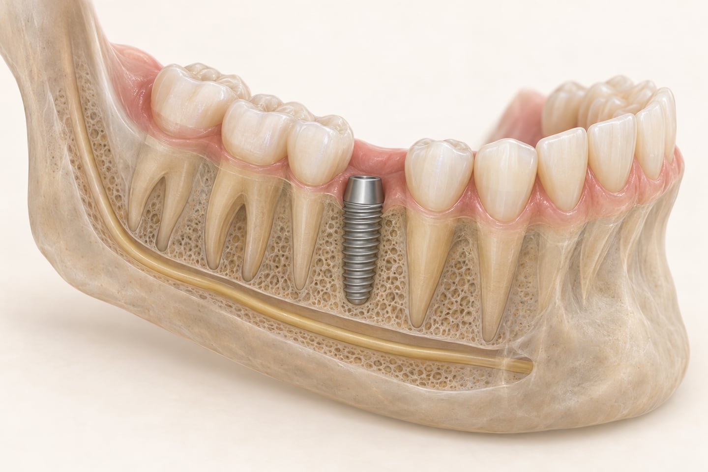
An implant is a post placed into your jawbone, and the bone is the one part nobody can see. A regular dental X-ray shows it as a flat picture. The three-dimensional scan that most implant plans include shows it as a shape that can be measured from any angle — and that is the difference between choosing where an implant goes and estimating.
What the scan is
The scan used for implant planning is usually cone beam computed tomography, shortened to CBCT. You sit or stand still while the machine rotates around your head once, capturing X-ray images from many angles that are assembled into a 3D model of the teeth, the jaw and the structures around them. The FDA lists dental implant planning among the uses of these systems. There is no tunnel to lie in and no injection; preparation is usually just removing glasses, earrings and any denture or partial.
Four things a flat X-ray can't tell you
A regular X-ray shows height. It cannot show how wide the ridge of bone is, and width is often what decides whether an implant fits at all. The scan answers the questions the plan depends on:
- Is there enough bone, in height and in width, to hold an implant of the right size?
- Where does the nerve that runs through the lower jaw sit, and how much bone is there above it?
- How close is the floor of the sinus to the roots of the upper back teeth?
- Is anything hidden that should be dealt with first — a leftover root tip, an infection at the end of a root, a cyst?
Those answers set the implant's size, angle and position, and whether something has to happen first: a bone graft, a sinus lift, or a different design altogether. Finding that out at the planning stage is the whole point. It is the difference between a plan and a surprise in the chair.
Planning from the finished tooth backward
The scan is not only about where the bone is. An implant has to sit where it can support a tooth that looks right, meets the opposite teeth properly and can be cleaned. So many plans start from the position of the final tooth and work backward to where the implant must go, and some turn the scan into a surgical guide — a template that seats over the teeth so the implant goes in exactly where it was planned. For a front tooth or a full arch, that planning is most of the work.
The radiation question
A CBCT scan uses X-rays, and it is worth knowing what that means. According to the FDA, dental CBCT exams generally deliver less radiation than other CT exams but typically more than conventional dental X-rays. The dose depends on the machine, the settings and how much of the head is scanned — a scan of one implant site covers far less than a scan of both jaws for a full-arch case. The FDA's advice is that the exam be used only when necessary, and that patients ask whether an equally useful exam with less or no radiation would do. For placing an implant near a nerve or a sinus, the scan usually is the exam that answers the question; for a routine checkup it is not. The ADA's page on dental X-rays, in the sources below, covers dental imaging more broadly.
Tell the office if you are or might be pregnant, and mention any recent scans from another office — if they can be sent over, nothing needs repeating.
What changes after the scan
The images are read alongside your exam, health history and goals and turned into a plan. For one missing tooth, that might be a simple yes, or a graft first. For several missing teeth it might be a choice between individual implants, a bridge on implants, or a denture held by implants. For a failing arch, it might be a full-arch plan. The implant procedure, step by step shows where the scan sits in the whole sequence.
If you have had a scan elsewhere
A CBCT scan is a file, not a film. If another office has taken one in the past year or so, ask them to export it — most systems produce a standard format that any planning software can read — and bring it to the consultation. Whether it can be reused depends on how much has changed in your mouth since and on how much of the jaw it covered, but it is worth asking before paying for a second one.
Ask where your scan will be taken
Not every office has a CBCT unit in the building; some refer patients to an imaging center and read the scan when it comes back. Neither is better, but it changes your visits, so ask up front. Ask, too, whether the scan will cover one site or both jaws, what it changed about the plan once it was read, and whether you can have a copy — useful if you ever want a second opinion. Request an implant consultation and put "imaging" in the notes so the first visit is planned around it.